
|
Welcome back! I’ve recently been interested in MRIs. Not because I’m injured or anything, but because of how it can make a picture of your brain structure. I think it would be so interesting to see what my brain looks like and compare it to others. In chapter 7 of Musicophilia, Sacks mentions a Harvard paper published in 1995 showing that the corpus callosum (band connecting the two brain hemispheres) is “enlarged in professional musicians”, and the planum temporale (in the auditory cortex) is enlarged in musicians with absolute pitch. A question of these enlargements was whether they were due to innate predisposition or musical training. Nevertheless, it’s evident that music is brain-altering! Playing an instrument involves many parts of the brain: the basal ganglia, cerebellum, and various areas of the cerebral cortex all show increased activity. Even just minutes of practice can show changes in the motor cortex. Below I have included a few interesting pictures/video of MRIs of musicians versus non-musicians, so you can see for yourself! Here is the link to a music video that I found pretty cool because it shows the creation of an MRI while the artist, Sivu, is singing: https://www.youtube.com/watch?v=_964dqQxQwY Video of taking fMRIs of Jennifer Koh, a professional violinist, to study the effects of reading, listening, and imagining music on the brain: https://today.duke.edu/2016/03/koh 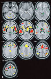 Brain while listening to music: 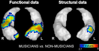 Comparison of brain images:
0 Comments
Leave a Reply. |
Archives
February 2021
Categories
All
|
 RSS Feed
